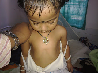
This is a case of Rickets which presented to our department with swelling of joints. The baby was 3 years old.
Clinical Images :
 Harrisons Groove
Upon evaluation of the case X-Ray was done on the wrist joints which revealed Cupping and Fraying of the metaphyseal ends of radius and ulna with widening of the metaphysis.

DISCUSSION :
Rickets is a softening of bones in children potentially leading to fractures and deformity. Rickets is among the most frequent childhood diseases in many developing countries. The predominant cause is a vitamin D deficiency, but lack of adequate calcium in the diet may also lead to rickets (cases of severe diarrhea and vomiting may be the cause of the deficiency). Although it can occur in adults, the majority of cases occur in children suffering from severe malnutrition, usually resulting from famine or starvation during the early stages of childhood.Osteomalacia is the term used to describe a similar condition occurring in adults, generally due to a deficiency of vitamin D.[1] The origin of the word "rickets" is probably from the Old English dialect word 'wrickken', to twist. The Greek derived word "rachitis" (ραχίτις, meaning "inflammation of the spine") was later adopted as the scientific term for rickets, due chiefly to the words' similarity in sound.
Signs and symptoms
Signs and symptoms of rickets include:
IMAGING FINDINGS
Plain radiograph findings include the following:
- Widening and cupping of the metaphyseal regions
- Fraying of the metaphysis
- Craniotabes
- Bowing of long bones
- Development of knock-knees, or genu valgum
- Development of scoliosis
- Impression of the sacrum and femora into the pelvis, leading to a triradiate configuration of the pelvis
- In healing rickets, the zones of provisional calcification become denser than the diaphysis. In addition, cupping of the metaphysis may become more apparent.
A useful mnemonic for remembering the findings of rickets is as follows:
- Reaction of the periosteum (may occur)
- Indistinct cortex
- Coarse trabeculation
- Knees, wrists, and ankles affected predominantly
- Epiphyseal plates, widened and irregular
- Tremendous metaphysis (cupping, fraying, splaying)
- Spur (metaphyseal)
Treatment and prevention
The treatment and prevention of rickets is known as
antirachitic.
[]
Diet and sunlight
Treatment involves increasing dietary intake of calcium, phosphates and vitamin D. Exposure to ultraviolet B light (sunshine when the sun is highest in the sky), cod liver oil, halibut live oil are all sources of vitamin D.
A sufficient amount of ultraviolet B light in sunlight each day and adequate supplies of calcium and phosphorus in the diet can prevent rickets. Darker-skinned babies need to be exposed longer to the
ultraviolet rays. The replacement of vitamin D has been proven to correct rickets using these methods of
ultraviolet light therapy and medicine.
Recommendations are for 400
international units (IU) of vitamin D a day for infants and children. Children who do not get adequate amounts of vitamin D are at increased risk of rickets. Vitamin D is essential for allowing the body to uptake calcium for use in proper bone calcification and maintenance.
[]Supplementation
Sufficient vitamin D levels can also be achieved through dietary supplementation and/or exposure to sunlight. Vitamin D
3 (
cholecalciferol) is the preferred form since it is more readily absorbed than vitamin D
2. Most
dermatologists recommend vitamin D supplementation as an alternative to unprotected ultraviolet exposure due to the increased risk of skin cancer associated with sun exposure. Note that in July in New York City at noon with the sun out, a white male in tee shirt and shorts will produce 20000
IU of Vitamin D from 20 minutes of non-sunscreen sun exposure.
[citation needed]According to the
American Academy of Pediatrics (AAP), infants who are breast-fed may not get enough vitamin D from breast milk alone. For this reason, the AAP recommends that infants who are exclusively breast-fed receive daily supplements of vitamin D from age 2 months until they start drinking at least 17 ounces of vitamin D-fortified milk or formula a day.
[2] This requirement for supplemental vitamin D is not a defect in the evolution of human breastmilk, but is instead a result of the modern-day infant's decreased exposure to sunlight (i.e. breast-fed infants who receive adequate sun exposure are less likely to develop rickets, though supplementation may still be indicated in the winter, depending on geographical latitude).


 Bow legs


