CROUZON SYNDROME
Treatment
TRACHEO-ESOPHAGEAL FISTULA - A case Report
This was a case of a 65 year old female who came to our department with dysphagia for Barium Swallow.
Upon doing the investigation, a tracheoesophageal fistula was detected and the patient was rushed to the casulaty for further management.
The patient had probably developed the fistula due to pressure necrosis by a tracheostomy tube as she had given history of being applied a trachestomy tube for quite some time.
X-Ray Images
DISCUSSION
A tracheoesophageal fistula is an abnormal connection (fistula) between the esophagus and the trachea. TEF is a common congenital abnormality, but when occurring late in life is usually the sequela of surgical procedures such as alaryngectomy.
Causes
Associations
Clinical presentation
Treatment
- Stricture, due to gastric acid erosion of the shortened esophagus.
- Leak of contents at the point of anastomosis.
- Recurrence of fistula.
RICKETS - A Case Report
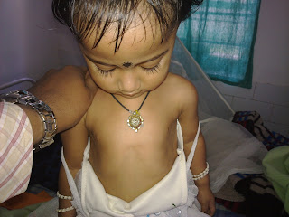
This is a case of Rickets which presented to our department with swelling of joints. The baby was 3 years old.
Clinical Images :

Signs and symptoms
- Bone pain or tenderness
- dental problems
- muscle weakness (rickety myopathy or "floppy baby syndrome" or "slinky baby" (where the baby is floppy or slinky-like)
- increased tendency for fractures (easily broken bones), especially greenstick fractures
- Skeletal deformity
- Toddlers: Bowed legs (genu varum)
- Older children: Knock-knees (genu valgum) or "windswept knees"
- Cranial, spinal, and pelvic deformities
- Growth disturbance
- Hypocalcemia (low level of calcium in the blood), and
- Tetany (uncontrolled muscle spasms all over the body).
- Craniotabes (soft skull)
- Costochondral swelling (aka "rickety rosary" or "rachitic rosary")
- Harrison's groove
- Double malleoli sign due to metaphyseal hyperplasia
- Widening of wrist raises early suspicion, it is due to metaphysial cartilage hyperplasia.
IMAGING FINDINGS
- Widening and cupping of the metaphyseal regions
- Fraying of the metaphysis
- Craniotabes
- Bowing of long bones
- Development of knock-knees, or genu valgum
- Development of scoliosis
- Impression of the sacrum and femora into the pelvis, leading to a triradiate configuration of the pelvis
- In healing rickets, the zones of provisional calcification become denser than the diaphysis. In addition, cupping of the metaphysis may become more apparent.
- Reaction of the periosteum (may occur)
- Indistinct cortex
- Coarse trabeculation
- Knees, wrists, and ankles affected predominantly
- Epiphyseal plates, widened and irregular
- Tremendous metaphysis (cupping, fraying, splaying)
- Spur (metaphyseal)
Treatment and prevention
[]
Diet and sunlight
Treatment involves increasing dietary intake of calcium, phosphates and vitamin D. Exposure to ultraviolet B light (sunshine when the sun is highest in the sky), cod liver oil, halibut live oil are all sources of vitamin D.
[]Supplementation
CHILAIDITI SYNDROME
Causes
Epidemiology
Synonyms
Retropharyngeal Abscess
INTRODUCTION
Retropharyngeal abscess (RPA) produces the symptoms of sore throat, fever, neck stiffness, and stridor. Retropharyngeal abscess occurs much less commonly today than in the past because of the widespread use of antibiotics for suppurative upper respiratory infections. Retropharyngeal abscess, once almost exclusively a disease of children, is observed with increasing frequency in adults. Retropharyngeal abscess poses a diagnostic challenge for the emergency physician because of its infrequent occurrence and variable presentation.
PATHOPHYSIOLOGY
- Aerobic organisms, such as beta-hemolytic streptococci and Staphylococcus aureus
- Anaerobic organisms, such as species of Bacteroides and Veillonella
- Gram-negative organisms, such as Haemophilus parainfluenzae and Bartonella henselae
- Widening of the retropharyngeal soft tissues is observed in 88% of patients with retropharyngeal abscess in a series that defined soft tissue swelling as more than 7 mm at C2 and more than 14 mm at C6. Most authors define retropharyngeal soft tissue swelling as more than 7 mm at C2 and more than 22 mm at C6; thus, lateral neck radiographs may be considerably less sensitive for detecting retropharyngeal abscess than this study indicates.
- Generally, the anteroposterior diameter of the prevertebral soft tissue space in children should not exceed that of the contiguous vertebral bodies.
- In addition to showing widening of the prevertebral space, the lateral neck radiograph rarely may show a gas-fluid level, gas in the tissues, or a foreign body.
Prehospital Care
- Supplemental oxygen and attention to upper airway patency are the essential components of prehospital care in patients with suspected retropharyngeal abscess.
- If a child exhibits respiratory distress, the sniffing position may be beneficial.
- Occasionally, endotracheal intubation or cricothyrotomy may be required if the patient exhibits signs of upper airway obstruction.
Emergency Department Care
- Airway management
- Apply supplemental oxygen. In young children, this can be completed in a nonthreatening way by letting the parent direct blow-by oxygen at the child's face.
- Endotracheal intubation may be required if the patient has signs of upper airway obstruction. It may be difficult because of upper airway swelling.
- Cricothyrotomy (surgical or needle) may be required in the patient with upper airway obstruction who cannot be intubated. Tracheostomy may be required for definitive airway management.
- Intravenous fluids are required if the patient is dehydrated because of fever and difficulty swallowing.


 Bow legs


