1

DIFFUSE PONTINE GLIOMA

HISTORY


A young child presented to our hospital with multiple cranial nerve palsies.


IMAGING

Intially a CT Scan was done for him followed by MRI.

CT SCAN


There was diffuse hypodensity seen involving the brainstem and a mass was suspected. The brainstem was also expanded.


MRI


The MRI revealed a diffuse non enhancing mass involving the pons mainly with expansion of the pons and compression of the surrounding cisterns.


It was diagnosed as a case of Diffuse Pontine Glioma.


DISCUSSION :

These tumours typically present in childhood (3 to 10 years of age) and make up 10 - 15% of all pediatric brain tumors. 

Presentation :
  • Multiple cranial nerve palsies
  • Signs of raised intracranial pressure. 
  • Cerebellar signs like ataxia, dysarthria, nystagmus and sleep apnoea.


Radiographic features

  • Enlarged pons 
  • Basilar artery  displaced anteriorly 
  • Flat floor of the fourth ventricle 
  • Obstructive Hydrocephalus  


Treatment and prognosis

  • Treatment commenced without histological confirmation. 
  • Chemotherapy only 
  • Children over 3 years of age - radiotherapy may be considered. 
  • Initial response can be dramatic and falsely reassuring.

Differential diagnoses
  • Rhombencephalitis
  • ADEM
  • NF1
  • Osmotic demyelination


0

GOSSYPIBOMA...

HISTORY
A 28 year old female presented to our hospital with a one day history of pain abdomen and vomiting.

She had got opearted about 10 days back - hysterectomy was done.

IMAGING


X-RAY


Plain X-Ray revealed - multiple air fluid levels suggesting intestinal obstruction and a vague mass in the left lumbar region with air foci within it.

CT - SCAN


CT Scan confirmed a heterogenous mass in the left lumbar region. It was seen separate from the bowel loops and was not showing enhancement and was diagnosed as Gossypiboma.

LAPAROTOMY was done for this patient and the surgical mop was retrieved. The patient came for follow up at 1 month and was having no symptoms subsequently.


DISCUSSION

Gossypiboma is the technical term for a surgical complications resulting from foreign materials, such as a surgical sponge , accidentally left inside a patient's body. The term "gossypiboma" is derived from the Latin gossypium (“cotton wool, ”) and the Swahili boma (place of concealment).

GI tract surgery and gynecologic surgery account for about 75% of reported gossypibomas
ž
žDifferential Diagnosis
Post-operative collection
Abscess
Hematoma


1

Maple Syrup Urine Disease (MSUD)

A typical case of MSUD


PRESENTATION: 

8 day old Term, AGA male baby, Parents had a non
 consanguinous marriage with no H/o unexplained death in family.
Baby presented with poor feeding, lethargy, persistent seizures, failure to thrive.
O/E baby was drowsy with tonic posturing of all limbs.


BIOCHEMICAL AND OTHER EVALUATION: 
Serum electrolytes, Calcium , Magnesium were normal.
Urine ketones positive
EEG Showed focal sharp wave activity over Left fronto-temporal areas.


CT revealed gross diffuse cerebral edema with involvement of the posterior fossa also


MRI FLAIR : Hyperintensities involving deep cerebellar regions with involvement of brainstem, thalami and corticospinal tracts.


MRI Diffusion : Restricted diffusion involving deep cerebellar regions with involvement of brainstem, thalami and corticospinal tracts.


1 week later Tandem Mass Spectroscopy revealed increased levels of branched chain amino acids (Valine, Leucineand Isoleucine) –confirming the diagnosis of MSUD


DISCUSSION:
Etiology: Autosomal recessive, Branched chain organic aciduria resulting from abnormalities of enzyme catabolism of branched chain amino acids. It leads to accumulation of BCAA and metabolites (neurotoxic). Accumulation of leucine in particular causes neurological symptoms. Increased Plasma isoleucine is associated with maple syrup odour.
Epidemiology : 1:850,000 general population, but can be as frequent as 1:170 in population isolates

Presentation:
Normal at birth , Presents after disease free interval, usually within the 4-7 days of life with poor feeding, vomiting, poor weight gain, increasing lethargy. Patients in crisis often smell like maple syrup
Maple syrup odor may be difficult to identify in first days of life unless urine soaked diaper is allowed to dry
Tandem mass spectrometry shortens diagnosis time
Mimic of sepsis: Acute encephalopathy, vomiting, seizures, neurological distress, lethargy, coma,
Plasma detection of alloisoleucine is diagnostic for MSUD
Typical EEG : "Comb-like-rhythms“


IMAGING FEATURES :
Classic appearing MSUD edema involving : Cerebellarwhite matter, brain stem, globuspallidus, thalamus, cerebral peduncles, corticospinaltracts (to cortex)
CT Findings
NECT : Diffuse edema NOT sparing brainstem and cerebellum
MR Findings
TlWI: decreased Signal intensity, margins may be sharp
T2WI : Generalized and MSUD edema
FLAIR: Generalized and MSUD edema
DWI : Marked restriction and decreased ADC (MSUD edema = intramyelinic)
MRS: Broad peak at chemical shift of 0.9 ppm
Ultrasonographic Findings
Increased Echogenicityof globuspallidi, periventricularwhite matter, and areas typically involved by MSUD edema


Treatment:
Acute "metabolic rescue" to reverse cerebral edema. May require hemodialysis during acute crisis to limit neurotoxicity/damage
Metabolically appropriate diet (protein-modified)minimizes severity
Prevent deficiencies of essential amino acids
Dietary therapy must be lifelong
Orthotopic liver transplantation increases availability of BCKD (rarely used)
Gene therapy experimental


0

Torsion testis - CASE STUDY

Hi guys.....posting after a really long time....
This is my first case of torsion testis.

Torsion testis


Findings :
Left testis was enlarged in size and heterogenous in apperance and most importantly NOT taking any vascularity. Right testis is there for comparison at the same blood flow settings.
Left cord was inflamed and hyperechoic in appearance and the vessels showed a 'whirlpool' appearance.


DISCUSSION

Introduction
Testicular torsion is when the spermatic cord to a testicle twists, cutting off the blood supply. The most common symptom is acute testicular pain and the most common underlying cause is a congenital malformation known as a "bell-clapper deformity". The diagnosis is often made clinically but if it is in doubt an ultrasound is helpful in ruling in or out the condition. Emergency diagnosis and treatment is required in order to save the viability of the testicle.

Signs and symptoms

Testicular torsion usually presents with an acute onset of diffuse testicular pain and tenderness of less than 6 hours of duration. There is often an absent or decreased cremasteric reflex. Many of the symptoms of testicular torsion are similar to the infection epididymitis.

Risk factors


Congenital

Conditions that allow the testicle to rotate predispose to torsion. A congenital malformation of the processus vaginalis known as the "bell-clapper deformity" accounts for 90% of all cases. In this condition, rather than the testes attaching posteriorly to the inner lining of the scrotum by the mesorchium, the mesorchium terminates early and the testis is free floating in the tunica vaginalis.


Size

A larger testicle either due to normal variation or a tumor increases the risk of torsion.


Temperature

Torsions are sometimes called "winter syndrome". This is because they often happen in winter, when it is cold outside. The scrotum of a man who has been lying in a warm bed is relaxed. When he arises, his scrotum is exposed to the colder room air. If the spermatic cord is twisted while the scrotum is loose, the sudden contraction that results from the abrupt temperature change can trap the testicle in that position. The result is a testicular torsion.


Diagnosis

Immediate testing for torsion is indicated when the onset of testicular pain is sudden and/or severe. In general a doppler ultrasound should be obtained only in low suspicion cases to rule out torsion while in those cases with a convincing history and physical exam immediate surgical detorsion (Derotation) is reasonable.

Clinical exam

Prehn's sign though a classic physical exam finding has not been found to be reliable in distinguishing torsion from other causes of testicular pain such asepididymitis. In cases of true torsion the cremasteric reflex is typically absent, the scrotum is generally not very swollen, and the affected testis may have a horizontal lie.


Imaging

doppler ultrasound scan of the scrotum is nearly 100% accurate at detecting torsion. It is identified by the absence of blood flow in the twisted testicle, which distinguishes the condition from epididymitis.


Pathophysiology

Torsion is due to a mechanical twisting process. It is also believed that torsion occurring during fetal development can lead to the so-called neonatal torsion orvanishing testis, and is one of the causes of an infant being born with monorchism (one testicle).


Treatment

With prompt diagnosis and treatment the testicle can be saved in a high number of cases. In some cases the testicle can untwist on its own or it can be manually untwisted, which can be attempted with pain relief as the guide for successful detorsion. Manual detorsion is successful in 26.5% to greater than 80% of patients based upon a number of reviewed studies.
Testicular torsion is a surgical emergency that needs immediate intervention. If treated within 6 hours, there is an excellent chance (90%) of saving the testicle. Within 12 hours the rate decreases to 50%, within 24 hours is 10%, and after 24 hours the rate approaches 0. Once the testicle is dead it must be removed to prevent gangrenous infection.


Epidemiology

Torsion is most frequent among adolescents with about 65% of cases presenting between 12 – 18 years of age. It occurs in about 1 in 160 males or 1 in 4000 males per year before 25 years of age.

1

CROUZON SYNDROME

CASE REPORT


This is a case report of an unusual case of a 12 year old child who presented to our department as a diagnosed case of Crouzon Syndrome with a post operative status of VP Shunt for reduction of Hydrocephalus. Hydrocephalus is common in crouzon syndrome because of Venous congestion.

Clinical Images


X-Ray showed : 


DISCUSSION
Crouzon syndrome is a genetic disorder known as a branchial arch syndrome. Specifically, this syndrome
affects the first branchial (or pharyngeal) arch, which is the precursor of the maxilla and mandible. Since the
branchial arches are important developmental features in a growing embryo, disturbances in their development
create lasting and widespread effects.
Causes
Associations with FGFR2 and FGFR3 have been identified.
Symptoms
As a very complicating result of the changes to the developing embryo, the symptoms are very pronounced features, especially in the facial areas. Low-set ears is a typical characteristic, as in all of the disorders which are called branchial arch syndromes. The reason for this abnormality is that ears on a fetus are much lower than those on an adult. During normal development, the ears "travel" upward on the head; however, in Crouzon patients, this pattern of development is disrupted. Ear canal malformations are extremely common, generally resulting in some hearing loss. In particularly severe cases, Ménière's disease may occur.
The most notable characteristic of Crouzon syndrome is cranial synostosis, but it usually presents asbrachycephaly, which results in the appearance of a short and broad head. Exophthalmos (bulging eyes due to shallow eye sockets after early fusion of surrounding bones), hypertelorism (greater than normal distance between the eyes), and psittichorhina (beak-like nose) are also symptoms. Additionally, a common occurrence is external strabismus, which can be thought of as opposite from the eye position found in Down syndrome. Lastly, hypoplastic maxilla (insufficient growth of the midface) results in relative mandibular prognathism (chin appears to protrude despite normal growth of mandible) and gives the effect of the patient having a concave face.
For reasons that are not entirely clear, most Crouzon patients also have noticeablyshorter humerus and femur bones, in proportion to the rest of their bodies, than members of the general population. A small percentage of Crouzon patients also have what is called "Type II" Crouzon syndrome, distinguished by partial syndactyly.
Diagnosis
Diagnosis of Crouzon syndrome usually can occur at birth by assessing the signs and symptoms of the baby. Further analysis, including radiographs, magnetic resonance imaging (MRI) scans, genetic testing, X-rays and CT scans can be used to confirm the diagnosis of Crouzon syndrome.
Incidence
Incidence of Crouzon syndrome is currently estimated to occur in 1 out of every 25,000 people out of the general population. 

Treatment

Surgery is typically used to prevent the closure of sutures of the skull from damaging the brain's development. Without surgery, blindness and mental retardation are typical outcomes. Craniofacial surgery is a discipline of plastic surgery. To move the orbits forward, plastic surgeons work with neurosurgeons to expose the skull and orbits and reshape the bone. To treat the midface deficiency, plastic surgeons can move the lower orbit and midface bones forward (this does not need neurosurgical assistance). For jaw surgery, either plastic surgeons have experience to perform these operations. It is rare to wear a custom-fitted helmet (or cranial band) for several months after surgery as that is only for single-suture "strip craniectomy" repair. Crouzon patients tend to have multiple sutures involved, most specifically bilateral coronal craniosynostoses, therefore an open operation is used rather than the strip craniectomy with helmeting.
Once treated for the cranial vault symptoms, Crouzon patients generally go on to live a normal lifespan.

0

TRACHEO-ESOPHAGEAL FISTULA - A case Report

This was a case of a 65 year old female who came to our department with dysphagia for Barium Swallow.

Upon doing the investigation, a tracheoesophageal fistula was detected and the patient was rushed to the casulaty for further management.
The patient had probably developed the fistula due to pressure necrosis by a tracheostomy tube as she had given history of being applied a trachestomy tube for quite some time.


X-Ray Images


DISCUSSION


A tracheoesophageal fistula is an abnormal connection (fistula) between the esophagus and the trachea. TEF is a common congenital abnormality, but when occurring late in life is usually the sequela of surgical procedures such as alaryngectomy.


Causes

Congenital TEF can arise due to failed fusion of the tracheoesophageal ridges during the third week of embryological development.
A fistula, from the Latin meaning ‘a pipe,’ is an abnormal connection running either between two tubes or between a tube and a surface. In tracheo-esophageal fistula it runs between the trachea and the esophagus. This connection may or may not have a central cavity; if it does, then food within the esophagus may pass into the trachea (and on to the lungs) or alternatively, air in the trachea may cross into the esophagus.
TEF can also occur due to pressure necrosis by a tracheostomy tube in apposition to a nasogastric tube (NGT).

Associations

Babies with TEF or esophageal atresia are unable to feed properly. Once diagnosed, prompt surgery is required to allow the baby to take in food. Few TEF children have problems after surgery, however a number develop feeding difficulties and chest problems. Some TEF babies are also born with other abnormalities, most commonly those described in VACTERL association - a group of anomalies which often occur together, including heart, kidney and limb deformities. 6% of babies with TEF also have a laryngeal cleft

Clinical presentation

Tracheoesophageal fistula is suggested in a newborn by copious salivation associated with chokingcoughing, vomiting, and cyanosis coincident with the onset of feeding. A fistula may also be a the cause of polyhydramnios while in utero.


Treatment


1

RICKETS - A Case Report

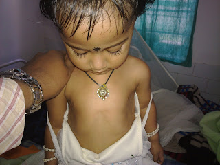
This is a case of Rickets which presented to our department with swelling of joints. The baby was 3 years old.

Clinical Images :

Harrisons Groove

Upon evaluation of the case X-Ray was done on the wrist joints which revealed Cupping and Fraying of the metaphyseal ends of radius and ulna with widening of the metaphysis.


DISCUSSION :

Rickets is a softening of bones in children potentially leading to fractures and deformity. Rickets is among the most frequent childhood diseases in many developing countries. The predominant cause is a vitamin D deficiency, but lack of adequate calcium in the diet may also lead to rickets (cases of severe diarrhea and vomiting may be the cause of the deficiency). Although it can occur in adults, the majority of cases occur in children suffering from severe malnutrition, usually resulting from famine or starvation during the early stages of childhood.Osteomalacia is the term used to describe a similar condition occurring in adults, generally due to a deficiency of vitamin D.[1] The origin of the word "rickets" is probably from the Old English dialect word 'wrickken', to twist. The Greek derived word "rachitis" (ραχίτις, meaning "inflammation of the spine") was later adopted as the scientific term for rickets, due chiefly to the words' similarity in sound.

Signs and symptoms

Signs and symptoms of rickets include:


IMAGING FINDINGS


Plain radiograph findings include the following:
  • Widening and cupping of the metaphyseal regions
  • Fraying of the metaphysis
  • Craniotabes
  • Bowing of long bones
  • Development of knock-knees, or genu valgum 
  • Development of scoliosis
  • Impression of the sacrum and femora into the pelvis, leading to a triradiate configuration of the pelvis
  • In healing rickets, the zones of provisional calcification become denser than the diaphysis. In addition, cupping of the metaphysis may become more apparent.
A useful mnemonic for remembering the findings of rickets is as follows:
  • Reaction of the periosteum (may occur)
  • Indistinct cortex
  • Coarse trabeculation
  • Knees, wrists, and ankles affected predominantly
  • Epiphyseal plates, widened and irregular
  • Tremendous metaphysis (cupping, fraying, splaying)
  • Spur (metaphyseal)


Treatment and prevention

The treatment and prevention of rickets is known as antirachitic.

[]


Diet and sunlight

Treatment involves increasing dietary intake of calcium, phosphates and vitamin D. Exposure to ultraviolet B light (sunshine when the sun is highest in the sky), cod liver oil, halibut live oil are all sources of vitamin D.

A sufficient amount of ultraviolet B light in sunlight each day and adequate supplies of calcium and phosphorus in the diet can prevent rickets. Darker-skinned babies need to be exposed longer to the ultraviolet rays. The replacement of vitamin D has been proven to correct rickets using these methods of ultraviolet light therapy and medicine.
Recommendations are for 400 international units (IU) of vitamin D a day for infants and children. Children who do not get adequate amounts of vitamin D are at increased risk of rickets. Vitamin D is essential for allowing the body to uptake calcium for use in proper bone calcification and maintenance.

[]Supplementation

Sufficient vitamin D levels can also be achieved through dietary supplementation and/or exposure to sunlight. Vitamin D3 (cholecalciferol) is the preferred form since it is more readily absorbed than vitamin D2. Most dermatologists recommend vitamin D supplementation as an alternative to unprotected ultraviolet exposure due to the increased risk of skin cancer associated with sun exposure. Note that in July in New York City at noon with the sun out, a white male in tee shirt and shorts will produce 20000 IU of Vitamin D from 20 minutes of non-sunscreen sun exposure.[citation needed]
According to the American Academy of Pediatrics (AAP), infants who are breast-fed may not get enough vitamin D from breast milk alone. For this reason, the AAP recommends that infants who are exclusively breast-fed receive daily supplements of vitamin D from age 2 months until they start drinking at least 17 ounces of vitamin D-fortified milk or formula a day.[2] This requirement for supplemental vitamin D is not a defect in the evolution of human breastmilk, but is instead a result of the modern-day infant's decreased exposure to sunlight (i.e. breast-fed infants who receive adequate sun exposure are less likely to develop rickets, though supplementation may still be indicated in the winter, depending on geographical latitude).